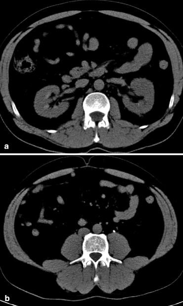

#Non obstructive left renal calculus trial
Small stones that do not require urgent urological intervention can be managed with symptomatic treatment and a trial of medical expulsive therapy to promote spontaneous passage. Diagnostics include spiral CT without contrast and/or ultrasound of the abdomen and pelvis to detect the stone, as well as urinalysis to assess for concomitant urinary tract infection ( UTI) and serum BUN and creatinine to evaluate kidney function.

Nephrolithiasis manifests as sudden-onset colicky flank pain that may radiate to the groin, testes, or labia, commonly called renal or ureteric colic, and it is usually associated with hematuria. Less common stones are composed of uric acid, struvite (due to infection with urease-producing bacteria), calcium phosphate, or cystine. Urinary stones are most commonly composed of calcium oxalate. Risk factors include low fluid intake and high- sodium, high- purine, low- potassium diets, which can raise the calcium, uric acid, and oxalate levels in the urine and thereby promote stone formation. Nephrolithiasis encompasses the formation of all types of urinary calculi in the kidney, which may be deposited along the entire urogenital tract, from the renal pelvis to the urethra.


 0 kommentar(er)
